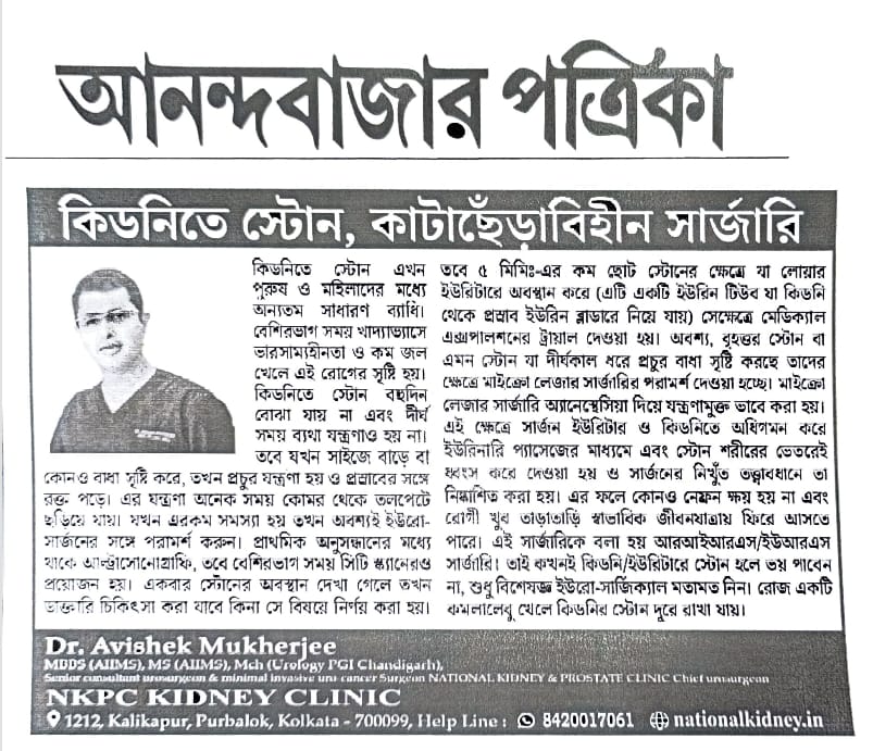
কিডনি এবং ইউরেটার স্টোন রোগ
-
National Kidney
- July 26, 2022
কিডনিতে পাথর
কিডনিতে পাথর আমাদের প্রভাবিত করে এমন একটি সাধারণ রোগ। এই পাথরগুলি মূলত ক্যালসিয়াম অক্সালেট জমা এবং স্ফটিক গঠন থেকে গঠিত হয়। ক্যালসিয়াম অক্সালেট স্ফটিকগুলি জলে দ্রবণীয় নয় এবং প্রস্রাবে সাইট্রিক অ্যাসিডের উপস্থিতি আমাদের এই স্ফটিকগুলিকে দ্রবীভূত করতে এবং তাদের নির্গত সাহায্য করে । কিন্তু কিছু লোকের সাইট্রিক অ্যাসিডের উৎপাদন কম থাকে এবং এই লোকেদের কিডনিতে তখন ক্যালসিয়াম অক্সালেট পাথর তৈরি হয় ।
ইউরিক অ্যাসিড স্ফটিকগুলি প্রায়শই প্রস্রাবে তৈরি হতে পারে এবং এগুলি ক্যালসিয়াম অক্সালেট পাথরের বিকাশের জন্য একটি নিডাস হিসাবে কাজ করতে পারে। তাই, যাদের রক্তে ইউরিক অ্যাসিডের মাত্রা বেশি তাদের ক্যালসিয়াম অক্সালেট পাথরও হতে পারে।
কদাচিৎ এই স্থবির প্রস্রাবে প্রস্রাবের স্থবিরতা এবং সংক্রমণ হতে পারে, যার ফলে সংক্রামক ম্যাট্রিক্স পাথর তৈরি হয়।
যখন প্রস্রাব দীর্ঘ সময়ের জন্য স্থবির হয়ে যায় তখন এই স্থবির প্রস্রাবে গৌণ পাথর হতে পারে, যেমনটি সাধারণত বাধাগ্রস্ত কিডনি এবং বাধাগ্রস্ত মূত্রথলিতে পাওয়া যায়।
পাথর রোগের প্রধান লক্ষণ কি কি?
প্রাথমিকভাবে কিডনিতে পাথর নীরব থাকে এবং কোনো উপসর্গ তৈরি করে না। কিন্তু যখন পাথর বড় হয়ে যায় বা প্রস্রাবপথে বাধা সৃষ্টি করে তখন রোগী প্রায়শই শরীরের পাশে ব্যথার সম্মুখীন হয় যা পুরুষদের তলপেটে বা টেস্টিস পর্যন্ত বিকিরণ করে।
প্রস্রাবের পথে পাথর আটকে গেলে লাল রঙের প্রস্রাব বা প্রস্রাবে রক্তও হতে পারে। কিছু পাথর প্রস্রাবে সংক্রমণ এবং দুর্গন্ধের সাথেও উপস্থিত হতে পারে। যেহেতু আজকাল নিয়মিত আল্ট্রাসনোগ্রাফি প্রায়ই সঞ্চালিত হয় তাই প্রায়শই নীরব পাথরও ঘটনাক্রমে সনাক্ত করা যায়।
পাথর রোগ নির্ণয়
পাথরের রোগ নির্ণয় করা হলে অবিলম্বে একজন প্রশিক্ষিত ইউরোসার্জন দ্বারা মূল্যায়ন করা উচিত। প্রাথমিকভাবে আল্ট্রাসনোগ্রাফি এবং প্রস্রাব এবং রক্ত পরীক্ষার মতো রুটিন পরীক্ষা করা হয়।
কিন্তু CT স্ক্যান হল পাথরের অবস্থান এবং আকার নির্ণয়ের সবচেয়ে গুরুত্বপূর্ণ হাতিয়ার এছাড়াও কিডনির কার্যকারিতা সার্জন দ্বারা সহজে এবং সঠিকভাবে মূল্যায়ন করা যায়।
তাই আজ পাথরের রোগ নির্ণয়ের জন্য CT স্ক্যান অপরিহার্য হয়ে উঠেছে।
চিকিৎসা ব্যবস্থাপনা
ওষুধের মাধ্যমে পাথর দ্রবীভূত করা যায় না এবং কিডনির অভ্যন্তরে অবস্থিত পাথর কোনো ওষুধের মাধ্যমে নির্গত হতে পারে না। তবে যেসব পাথরের আকার 5 মিমি-এর চেয়ে ছোট এবং মূত্রনালীতে চলে গেছে, তাদের চিকিৎসা পরীক্ষার মাধ্যমে চিকিৎসা করা যেতে পারে। ইউরেটার হল সরু নল যা প্রতিটি কিডনি থেকে মূত্রাশয় পর্যন্ত প্রস্রাব বহন করে। মূত্রাশয় যেখানে প্রবেশ করে তার নিচের অংশে ইউরেটার সবচেয়ে সরু। তাই মূত্রনালীর এই অংশে পাথর সবচেয়ে খারাপভাবে প্রভাবিত হয়।
আমাদের কাছে ট্যামসুলোসিন নামে একটি ওষুধ রয়েছে, যা ইউরেটারের নীচের বেশিরভাগ অংশের পেশীগুলিকে শিথিল করতে পারে। অতএব, যখন পাথর 5 মিমি-এর চেয়ে ছোট হয় এবং মূত্রনালীর সর্বনিম্ন অংশে অবস্থিত, তখন একটি মেডিকেল ট্রায়ালের চেষ্টা করা যেতে পারে।
যাইহোক, এটা খুবই গুরুত্বপূর্ণ যে, রেনাল ফাংশন ভাল এবং রোগীর কোন সংক্রমণ বা তীব্র ব্যথা নেই। তবে কিডনি প্রসারিত এবং সংক্রামিত হলে এবং বা গুরুতর ব্যথা হলে, মেডিকেল ট্রায়াল চেষ্টা করা উচিত নয়।
একবার আমরা একটি মেডিক্যাল ট্রায়াল শুরু করলে রোগীকে ঘনিষ্ঠভাবে অনুসরণ করা হয়। যদি রোগীর তীব্র ব্যথা বা জ্বর হয় তবে আমাদের অবিলম্বে অস্ত্রোপচারের কথা ভাবতে হবে ।
মেডিক্যাল ট্রায়াল মাত্র দুই সপ্তাহের জন্য চালিয়ে যেতে পারে, যদি পাথর দুই সপ্তাহের মধ্যে পাস না হয়, তাহলে আমাদের ব্যবস্থাপনাটিকে মাইক্রোসার্জিক্যাল কেয়ারে রূপান্তর করতে হবে। অতএব, দুই সপ্তাহের চিকিৎসা পরীক্ষার পর আমরা রোগীকে পাথর কেটে গেছে কি না তা নির্ণয়ের জন্য একটি সিটি স্ক্যান করার পরামর্শ দিই।
যদি পাথর দুই সপ্তাহ পার না হয় তবে এন্ডো-সার্জিক্যাল ব্যবস্থাপনায় রূপান্তর করার পরামর্শ দেওয়া হয়।
- 6 মিমি থেকে কম 1টি ছোট মূত্রনালীর পাথর চিকিৎসা পরীক্ষার জন্য সবচেয়ে উপযুক্ত
- নিম্ন মূত্রনালীতে অবস্থিত 2টি পাথর চিকিৎসা পরীক্ষার জন্য সবচেয়ে উপযুক্ত
- মেডিকেল ট্রায়াল সম্ভব যখন কিডনি ভাল কাজ করে এবং রোগীর কোন সংক্রমণ থাকে না এবং সামান্য বা কোন ব্যথা হয় না
- টি মেডিকেল ট্রায়াল দুই সপ্তাহের জন্য করা যেতে পারে এবং এর বেশি নয় কারণ দুই সপ্তাহ পর কিডনি স্থায়ীভাবে ক্ষতিগ্রস্থ হতে পারে
পাথরের রোগে কখন অস্ত্রোপচারের চিকিৎসা অপরিহার্য?
যখনই কিডনিতে পাথর 6 মিমি-এর চেয়ে বড় হয় তখন এন্ডো-সার্জারিভাবে চিকিত্সা করা ভাল কারণ কিডনিতে পাথরের জন্য কোনও চিকিৎসা ব্যবস্থা উপলব্ধ নেই। যাইহোক, ইউরেটার পাথরের ক্ষেত্রে, যেগুলি 6 মিলিমিটারের চেয়ে বড়, বা যেগুলি চিকিত্সা ব্যবস্থাপনার মাধ্যমে পাস করার সম্ভাবনা নেই তাদের লেজার এন্ডো-সার্জারি দ্বারা সর্বোত্তম চিকিত্সা করা হয়। এছাড়াও যদি মূত্রনালীর পাথরের ক্ষেত্রে মেডিকেল ট্রায়াল ব্যর্থ হয় তবে আমাদের অবিলম্বে অস্ত্রোপচারের যত্ন নেওয়া উচিত।
6 মিমি থেকে বড় কিডনিতে 1টি পাথর এন্ডোসার্জারির মাধ্যমে সর্বোত্তম চিকিত্সা করা হয়
6 মিমি থেকে বড় 2টি ইউরেটার পাথর অস্ত্রোপচারের মাধ্যমে সর্বোত্তম চিকিত্সা করা হয়
3 যে সমস্ত মূত্রনালীর পাথর চিকিৎসা ব্যবস্থাপনার জন্য উপযুক্ত নয় বা চিকিৎসা পরীক্ষা ব্যর্থ হয়েছে সেগুলোও অস্ত্রোপচারের জন্য বেছে নেওয়া হয়।
স্টোন এন্ডো সার্জারি কি?
আগে ওপেন সার্জারির মাধ্যমে সব পাথরের অপারেশন করা হতো। তবে এই কৌশলগুলি বেশিরভাগই অপ্রচলিত হয়ে গেছে এবং এখন পাথরের রোগের ক্ষেত্রে খোলা অস্ত্রোপচারের খুব কমই প্রয়োজন হয়। আজকাল আমরা এন্ডো-সার্জারি ব্যবহার করি, যার মানে আমরা মূত্রপথে প্রবেশের জন্য খুব সূক্ষ্ম টেলিস্কোপ ব্যবহার করি, এগুলি প্রস্রাবের প্রবাহের স্বাভাবিক পথ। প্যাসেজের ভিতরে পাথরটি স্থানীয়করণ করা হয় এবং তারপর শক্তি ব্যবহার করে পাথরটি খণ্ডিত হয় এবং তারপরে সরানো হয়।
অতএব, রোগীর ট্রমা খুব সীমিত এবং রোগী খুব কম ব্যথা নিয়ে সেরে উঠতে পারে।
অন্যদিকে লেজারগুলি একটি প্লাজমা বুদবুদ তৈরি করে যা পাথরকে ধ্বংস করে এবং সেইজন্য অবশিষ্ট পাথরের সম্ভাবনা খুবই সীমিত, এছাড়াও পাথরটিকে পিছনে ঠেলে দেওয়া হয় না, তবে এটি যেখানে রাখা হয়েছে ঠিক সেখানেই ধ্বংস করা হয়।
তাই আজ যে কোনও বড় পাথর কেন্দ্রে লেজারগুলি পাথর রোগের ব্যবস্থাপনায় অপরিহার্য হয়ে উঠেছে।
পাথর রোগে লেজার
যখন সার্জন প্রস্রাবের ট্র্যাক্টে প্রবেশ করে এবং তারপর পাথরটি কল্পনা করতে পারে, তখন পাথরটি সাধারণত শক্ত হয় এবং প্রায় কখনই অক্ষত অবস্থায় বের করা যায় না। অতএব, অস্ত্রোপচারকে পাথরটি ধ্বংস বা খণ্ডিত করার জন্য শক্তির কিছু উৎস ব্যবহার করতে হবে। এটি পাথরটিকে ছোট ছোট টুকরোতে পরিণত করার অনুমতি দেয় যা পরে অপসারণ করা যেতে পারে বা তারা নিজেরাই বেরিয়ে যেতে পারে।
আগে আমরা বায়ুসংক্রান্ত হাতুড়ি ব্যবহার করতাম, যা পাথর ভাঙার জন্য মাইক্রো-হ্যামার। কিন্তু এই হাতুড়িগুলি প্রায়শই বড় টুকরো তৈরি করে, যা মূত্রনালীতে ফিরে থাকতে পারে যার ফলে অবশিষ্ট পাথরের বোঝা তৈরি হয়।
শক ওয়েভ লিথোট্রিপসি কেন আজকাল ব্যবহার করা হয় না?
ইএসডব্লিউএল বা এক্সট্রাকর্পোরিয়াল শক ওয়েভ লিথোট্রিপসি অতীতে খুব জনপ্রিয় ছিল। এই প্রজন্মের শক ওয়েভ, যা শরীরে প্রবেশ করতে পারে এবং শক্ত পাথর টুকরো টুকরো করে দিতে পারে এবং নরম কিডনিকে বাঁচাতে পারে। তবে এটি এখন প্রায় অপ্রচলিত হয়ে গেছে, যেহেতু শক তরঙ্গগুলি প্রায়শই বড় টুকরো তৈরি করে এবং প্রায়শই পাথরটি পুরোপুরি পরিষ্কার হয় না।
পাথরের সম্পূর্ণ ক্লিয়ারেন্সের জন্য এটি একাধিক সেশনও নিতে পারে এবং অনেক ক্ষেত্রে একাধিক সেশনের পরেও পাথরটি পুরোপুরি পরিষ্কার নাও হতে পারে।
শক ওয়েভের কারণে মূত্রনালী এবং কিডনির ক্ষতি হওয়ার প্রশ্নও রয়েছে এবং পাথরের বড় ধারালো এবং শক্ত টুকরোগুলি মূত্রনালী বা কিডনিতে প্রভাবিত হচ্ছে।
1 ESWL এর ফলে প্রায়শই পাথরের বড় টুকরো হয়
একাধিক সেশনের পরেও 2 পাথর ক্লিয়ারেন্স অসম্পূর্ণ হতে পারে
3 eswl এর ফলে কিডনি বা মূত্রনালী ক্ষতিগ্রস্ত হতে পারে
4 এটি এখন খুব কমই ব্যবহৃত হয় যেহেতু লেজারগুলি পাথরের রোগের ব্যবস্থাপনাকে সম্পূর্ণরূপে পরিবর্তন করেছে
ইউরেটেরোস্কোপি এবং লেজার লিথোট্রিপসি
যখন পাথরটি ইউরেটারে থাকে তখন আমরা পাথরটি ধ্বংস করার জন্য ইউরেটেরোস্কোপি এবং লেজার লিথোট্রিপসি করি। ইউরেটার হল সেই টিউব যা কিডনি থেকে মূত্রাশয় পর্যন্ত প্রস্রাব বহন করে এবং ইউরেটারোস্কোপ হল এন্ডোস্কোপ যা আমাদের মূত্রনালীতে প্রবেশ করতে এবং পাথরের উপর সঠিকভাবে লেজারকে ফোকাস করতে দেয়।
এই প্রক্রিয়াটি প্রাকৃতিক প্রস্রাবের মাধ্যমে সঞ্চালিত হয় এবং তাই এই পদ্ধতির জন্য কোন কাটা বা ছেদ প্রয়োজন নেই। এই পদ্ধতিটি সাধারণত মেরুদণ্ডের অ্যানেস্থেশিয়ার অধীনে সঞ্চালিত হয়, যদি না সাধারণ অ্যানেশেসিয়া দাবি করার জন্য অন্য কিছু কারণ থাকে।
একবার পাথর ফোকাস হয়ে গেলে আমরা উন্নত হলমিয়াম লেজার ব্যবহার করি। পাথরের উপর নিবদ্ধ লেজার শক্তি পাথরকে ধ্বংস করতে পারে এবং তাই রোগীকে পাথর মুক্ত করা যেতে পারে।
1 ইউরেটার হল একটি টিউব যা কিডনি থেকে মূত্রাশয় পর্যন্ত প্রস্রাব নিষ্কাশন করে
2 ureteroscopy হল এন্ডোস্কোপিক পদ্ধতি যা প্রাকৃতিক মূত্রপথের মাধ্যমে মূত্রনালীতে প্রবেশ করে
3 আমরা পাথর ধ্বংস করতে লেজার ব্যবহার করি
4 সাধারণত এটি মেরুদণ্ডের অ্যানেস্থেশিয়ার অধীনে সঞ্চালিত হয় এবং রোগীকে এক থেকে দুই রাত থাকার পরে ছেড়ে দেওয়া যেতে পারে
RIRS
রেট্রোগ্রেড ইন্ট্রারেনাল সার্জারি, বা RIRS হল মূত্রপথের মাধ্যমে প্রবর্তিত নমনীয় ইউরেটেরো-রেনোস্কোপ দ্বারা কিডনি থেকে পাথর অপসারণ।
নমনীয় uretero-renoscope হল একটি দীর্ঘ সরু এবং নমনীয় এন্ডোস্কোপ। এটি অ্যানেস্থেশিয়ার অধীনে প্রাকৃতিক প্রস্রাব পথের মাধ্যমে স্থাপন করা হয়। সার্জন কিডনির মধ্যে আলোচনা করতে সক্ষম হয় যেখান থেকে তিনি ক্যালিসিয়াল সিস্টেমে প্রবেশ করেন। তারপর একবার পাথরটি দৃশ্যমান হয়ে গেলে পাথরটি ধ্বংস করতে একটি লেজার ব্যবহার করা হয়।
এই পদ্ধতিটি 10 মিমি থেকে ছোট এবং কিডনির মধ্যে অবস্থিত পাথরের জন্য সঞ্চালিত হয়।
1 রেট্রোগ্রেড ইন্ট্রা রেনাল সার্জারি 6-10 মিমি আকারের পাথরের জন্য বোঝানো হয়
2 অস্ত্রোপচার এনেস্থেসিয়া অধীনে সঞ্চালিত হয়
3 প্রক্রিয়াটি প্রাকৃতিক প্রস্রাবের মাধ্যমে সঞ্চালিত হয় এবং প্রবেশের জন্য কোন ছেদনের প্রয়োজন হয় না
4 পাথর সম্পূর্ণরূপে খণ্ডিত, কিন্তু ক্লিয়ারেন্স একটি সিটি স্ক্যান দ্বারা দুই মাস পরে নিশ্চিত করা হয়.
MINI perc
এটি ইউরো সার্জনের অস্ত্রাগারের সর্বশেষ সংযোজনগুলির মধ্যে একটি। এখানে এনেস্থেশিয়ার অধীনে পদ্ধতিটি সঞ্চালিত হয়। সার্জন একটি 5 মিমি বা তার চেয়ে ছোট ছেদ রাখে এবং খুব উন্নত মাইক্রোসার্জারি টেলিস্কোপ ব্যবহার করে কিডনিতে প্রবেশ করে এবং লেজার সার্জারি ব্যবহার করে পাথর অপসারণ করে।
যেহেতু ব্যবহৃত টেলিস্কোপগুলি খুব সূক্ষ্ম তাই কিডনি ন্যূনতম ট্রমা বজায় রাখে এবং ফলাফল খুব ভাল হয়।রোগী খুব দ্রুত কাজে ফিরতে পারে।
- 10 মিমি থেকে বড় কিডনি পাথরের জন্য এটি আদর্শ কৌশল
- 2 যেহেতু ব্যবহৃত টেলিস্কোপটি খুব সূক্ষ্ম তাই কিডনিতে আঘাত খুব কম
- রোগীর ন্যূনতম ব্যথা হয় এবং কাজে ফিরতে পারেন খুব তাড়াতাড়ি
PCNL
এটি কিডনিতে অনেক বড় এবং অবস্থিত পাথর অপসারণের জন্য সময় পরীক্ষিত এবং নির্ভরযোগ্য কৌশল। এই পাথরগুলি প্রচলিত বড় ক্যালিবার নেফ্রোস্কোপ দ্বারা অপসারণ করা হয় যেখানে এমনকি খুব বড় স্টাগহর্ন পাথরগুলিও সরানো যেতে পারে।
এই রোগীরা দুই থেকে চার দিন হাসপাতালে থাকে এবং দুই সপ্তাহ পরে কাজে ফিরতে পারে।
1 এটি 20 মিমি-এর বেশি আকারের খুব বড় রেনাল পাথরের জন্য ডিজাইন করা হয়েছে
2 এখানে আমরা এই পাথর অপসারণের জন্য একটি বড় ক্যালিবার নেফ্রোস্কোপ ব্যবহার করি
3 রোগী অস্ত্রোপচারের দুই সপ্তাহ পরে কাজে ফিরতে পারেন
এসব মামলা ফলো আপ করুন
সাধারণত, রোগীদের এক মাস পর নতুন সিটি স্ক্যান করার পরামর্শ দেওয়া হয়। পাথরের সম্পূর্ণ ক্লিয়ারেন্স নিশ্চিত করতে। যদি একটি স্টেন্ট স্থাপন করা হয় তাও সরানো হয়। এই তারিখের পরে রোগী স্বাভাবিক কাজ এবং জীবন পুনরায় শুরু করতে পারেন।
পাথর রোগে পথ্য
প্রস্রাবে কম সাইট্রিক অ্যাসিডের উপস্থিতির কারণে প্রায়শই কিডনিতে পাথর তৈরি হয়। তাই এই রোগীদের প্রতিদিন ও সারাজীবন একটি করে মোসাম্বি বা কমলা খেতে বলা উচিত। যেহেতু এই সাইট্রাস ফলগুলিতে উচ্চ মাত্রার সাইট্রিক অ্যাসিড থাকে তাই এই রোগীদের আবার পাথর হওয়ার সম্ভাবনা থাকে না, যতক্ষণ না সাইট্রাস ফল প্রতিদিন খাওয়া হয়। যেহেতু লেবুতে প্রচুর পরিমাণে ভিটামিন সি রয়েছে এবং এটি রেনাল স্টোন গঠনের সম্ভাবনা বাড়িয়ে তুলতে পারে তাই সাধারণ লেবু, নিম্বু এড়িয়ে চলা ভাল।
Prostate disease medical management versus laser surgery
-
National Kidney
- July 21, 2022
Prostate is the gland which is located below the urinary bladder in men. It produces semen and therefore is very important for the purpose of reproduction. With increase in age, it enlarges and very often can result in obstruction to the urine passage. These patients very often feel that the flow of the urine has reduced, also they may need to pass urine very frequently during day and night.
If one is facing these problems, please consult an uro-surgeon immediately. The uro-surgeon will generally examine the patient and then will advise, for a few routine tests. These tests include a urine culture and routine test, blood for hemoglobin, creatinine, sugar and PSA. Also, a urine flow test and an ultrasound evaluation is routinely performed. Based on these tests the surgeon is able to assess the extent of the problem and therefore the plan of care is also decided.
When the urine culture is sterile, and the PSA is less than 2, and the urine flow is more than 15ml/sec and the residual urine is less than 100ml, then most such patients are best managed by medication. These medications are like blood pressure medication and can be used for years, under the supervision of the uro-surgeon. Generally, these patients are called for twice per year check up.
However in case the urine culture shows infection, and the PSA is more than 2 and the urine flow is less than 10ml/sec , and the residual urine is more than 100ml, then the patient has more serious obstruction which needs care by microsurgery. These patients are then called for cystoscopy, which is a day care procedure performed under local anesthesia to check the internal anatomy of the prostate. These patients are better cared for by laser surgery of the prostate.
Laser surgery is a no cut, natural passage surgery. Which is performed using laser therefore the chance of bleed, infection and residual prostate is very small. And the patient can recover very soon from the surgery.
Therefore if you are suffering from urinary symptoms please consult an uro-surgeon soon.
KIDNEY AND URETER STONE DISEASE
-
National Kidney
- July 21, 2022
Kidney stones
Kidney stones are one of the commonest diseases affecting us. These stones are formed mainly from calcium oxalate deposition and crystal formation. Calcium oxalate crystals are not water soluble and the presence of citric acid in the urine allow us to dissolve these crystals and excrete them. But some people have lesser production of citric acid and these people then develop calcium oxalate stones.
Uric acid crystals can often form in the urine and these can serve as a nidus for the development of the calcium oxalate stones. Therefore, patients who have higher uric acid levels in the blood can also develop calcium oxalate stones.
Rarely one can have stagnation of urine and infection in this stagnant urine, which results in production of infective matrix stones.
When urine gets stagnant for a long time then one can develop secondary stones in this stagnant urine, as is typically found in obstructed kidney and obstructed urinary bladder.
What are the main symptoms of stone disease?
Initially the kidney stones are silent and don’t produce any symptoms. But as the stones become larger or start obstructing the urine passage then the patient often encounters pain in the side of the body which radiates down to the lower abdomen or to the testis in men.
In case the stone gets stuck in the urine passage it can also cause red colored urine or blood in the urine. Some stones may also present with infection and foul smell in the urine.
Since nowadays routine ultrasonography is often performed therefore often silent stones can also be detected incidentally .
Diagnosis of stone disease
Stone disease when diagnosed should be immediately evaluated by a trained uro-surgeon. Initially routine tests are performed like ultra-sonography and urine and blood tests.
But CT scan is the most important tool to assess the location and the size of the stone also the kidney function can be easily and accurately assessed by the surgeon.
Therefore today ct scan has become indispensable in assessment of stone disease.
Medical management
Stones cannot be dissolved by medication and stones located inside the kidney cannot be excreted by any medication. However stones which are smaller than 5mm in size and have passed into the ureter, can be treated with a medical trial. Ureter is the narrow tube which carries the urine from each of the kidneys to the bladder. The ureter is narrowest in the lowest part where it enters the bladder. Therefore the stones get most badly impacted in this part of the ureter.
We have a medicine called tamsulosin, which can relax the muscles of the lower most part of the ureter. Therefore, when the stones are smaller than 5mm, and located in the lowest part of the ureter, a medical trial can be attempted.
However, it is very important that, the renal function is good and the patient has no infection or severe pain. If however the kidney is dilated and infected and or there is severe pain, then medical trial should not be tried.
Once we start a medical trial then the patient is kept in close follow up. If the patient develops severe pain or fever then we have to proceed to surgical care immediately.
Medical trial can be continued for only two weeks, if the stone has not passed in two weeks, then we need to convert the management to microsurgical care. Therefore, after two weeks of medical trial we advise the patient to undergo a ct scan to assess whether the stone has passed or not.
If the stone has not passed by two week then it is advisable to convert to endo-surgical management.
1 small ureteral stones less than 6mm are best suited for medical trial
2 stone located in the lower ureter are best suited for medical trial
3 medical trial is possible when the kidney has good function and the patient has no infection and little or no pain
4 medical trial can be carried out for two weeks and not more since the kidney can sustain permanent damage after two weeks.
When is surgical treatment essential in stone disease?
Whenever kidney stones are larger than 6mm it is best to treat them endo-surgically since no medical management is available for renal stones. However, in the case of ureter stones, those which are larger than 6mm, or those which are unlikely to pass with medical management are best treated with laser endo-surgery. Also if medical trial has failed in case of ureteral stones then also we should immediately opt for surgical care.
1 stone in the kidney larger than 6mm are best treated with endosurgery
2 ureter stone larger than 6mm are best treated with surgery
3 those ureteral stone which are not suitable for medical management or medical trial has failed are also selected for surgical care.
trial has failed are also selected for surgical care.
What is stone endo-surgery?
Earlier all stone were operated by open surgery. However these techniques have mostly become obsolete and now open surgery is hardly ever needed in cases of stone disease. Nowadays we use endo-surgery, which means we use very fine telescopes to enter the urinary passage, these being the natural passage of urine flow. Inside the passage the stone is localized and then using energy the stone is fragmented and then removed.
Therefore, the trauma to the patient is very limited and the patient can recover with very less pain.
Lasers in stone disease
When the surgeon enters the urine tract and then can visualize the stone, the stone is generally hard and can almost never be pulled out intact. Therefore, the surgeon must use some source of energy to destroy or fragment the stone. This allows the stone to be reduced to small fragments which can then be removed or they can pass out by themselves.
Earlier we used pneumatic hammers, which are micro-hammers to break the stones. But these hammers very often created larger fragments, which could remain back in the urinary system resulting in residual stone burden.
Lasers on the other hand create a plasma bubble which destroys the stone and therefore the chance of residual stones is very limited, also the stone is not pushed back, but destroyed exactly at the site where it is lodged.
Therefore today in any major stone center lasers have become indispensable in the management of stone disease.
Why is shock wave lithotripsy not used nowadays?
ESWL or extracorporeal shock wave lithotripsy was very popular in the past. This entailed generation of shock waves, which can pass into the body and fragment the hard stones, while sparing the softer kidney. However this has become almost obsolete now, since the shock waves very often create large fragments, and very often the stone is not cleared completely.
It also may take multiple sessions for the complete clearance of the stone, and in many cases even after multiple sessions the stone may not be cleared completely.
There is also the question of damage to the ureter and the kidney due to the shock waves and also the large sharp and hard fragments of the stone getting impacted into the ureter or the kidney.
1 ESWL results very often in larger fragments of the stone
2 stone clearance may be incomplete even after multiple sessions
3 eswl may result in kidney or ureter damage
4 it is hardly used now since lasers have changed the management of stone disease completely
Ureteroscopy and laser lithotripsy
When the stone is located in the ureter then we perform ureteroscopy and laser lithotripsy to destroy the stone. Ureter is the tube which carries the urine from the kidney to the bladder, and ureteroscope is the endoscope which allows us to access the ureter and focus the laser accurately on the stone.
This procedure is performed via the natural urine passage and therefore there is no cut or incision needed for this procedure. This procedure is generally performed under spinal anesthesia, unless there are some other factors demanding general anesthesia.
Once the stone is focused then we use the advanced holmium laser. The laser energy focused on the stone can destroy the stone and therefore the patient can be rendered stone free.
1 ureter is the tube which drains the urine from the kidney to the bladder
2 ureteroscopy is the endoscopic procedure to access the ureter via the natural urine passage
3 we use laser to destroy the stone
4 generally this is performed under spinal anesthesia and the patient can be discharged after one to two nights stay.
RIRS
Retrograde intrarenal surgery, or RIRS, is the removal of stones from the kidney, by a flexible uretero-renoscope, introduced via the urine passage.
The flexible uretero-renoscope is a long narrow and flexible endoscope. It is placed via the natural urine passage, under anesthesia. The surgeon is able to negotiate to within the kidney from where he enters the calyceal system. Then once the stone is visualized then a laser is used to destroy the stone.
This procedure is performed for stones which are smaller than 10mm and located within the kidney.
1 retrograde intra renal surgery is meant for stones 6-10 mm in size
2 the surgery is performed under anesthesia
3 the procedure is performed via the natural urine passage and no incision is needed for the access
4 the stones are completely fragmented, but the clearance is confirmed after two month by a CT scan.
MINI PERC
This is one of the latest additions to the arsenal of the uro surgeon. Herein under anesthesia the procedure is performed. The surgeon places a 5mm or smaller incision and using very advanced microsurgery telescope enters the kidney and removes the stone using laser surgery.
Since the telescopes used are very fine therefore the kidney sustains mnimum trauma and the results are very good.
The patient can return to work very fast.
1 for kidney stones larger than 10mm this is the ideal technique
2 since the telescope used is very fine therefore the trauma to the kidney is very minimal
3 patient has minimum pain and can return to work very soon .
PCNL
This is time tested and reliable technique for removal of stones which are very large and located in the kidney. These stones are removed by conventional large caliber nephroscope where in even very large staghorn stones can be removed.
These patients stay in the hospital for two to four days and can return to work after two week.
1 this is designed for very large renal stones more than 20mm in size
2 here we use a large caliber nephroscope for removal of these stones
3 the patient can return to work after two weeks of the surgery
Follow up of these cases
Generally, the patients are advised to review after one month with a fresh ct scan. To confirm the complete clearance of the stones. If a stent has been placed it is also removed. After this date the patient can resume normal work and life.
Diet in stone disease
The kidney stones are most often formed due to presence of less citric acid in the urine. Therefore, these patients should be asked to consume one mousambi or orange daily and lifelong. Since these citrus fruits contain high levels of citric acid therefore these patients are not likely to form stones again, as long as the citrus fruit is consumed daily. Since lemon contains large quantities of vitamin c and this can increase the chance of renal stone formation therefore the common lemon, nimbu is best avoided.
কিভাবে প্রোস্টেট ক্যান্সার থেকে নিজেকে বাঁচবেন ?
-
National Kidney
- July 8, 2022
প্রস্টেট কি?
প্রোস্টেট হল একটি গ্রন্থি যা শুধুমাত্র পুরুষদের মধ্যে থাকে, যেমন মহিলাদের জরায়ু থাকে। প্রোস্টেট মূত্রথলির গোড়ায় অবস্থিত। এটি বীর্য উৎপন্ন করে এবং তাই এটি একটি প্রজনন অঙ্গ। 40 বছর বয়সের পরে সৌম্য বৃদ্ধির কারণে প্রোস্টেট বড় হতে পারে, তবে কিছু ক্ষেত্রে গ্রন্থিটিতে ক্যান্সার হতে পারে।
প্রোস্টেট ক্যান্সারের কারণ কি?
প্রোস্টেট ক্যান্সার গঠনের সঠিক কারণ জানা যায়নি। যাইহোক, প্রোস্টেট ক্যান্সারের বিকাশের জন্য পারিবারিক ইতিহাস একটি শক্তিশালী ভবিষ্যদ্বাণীমূলক কারণ । অতএব, যদি একটি পরিবারে প্রোস্টেট ক্যান্সারের ইতিহাস থাকে তবে 40 বছর বয়সের পরে পরিবারের সমস্ত পুরুষদের বছরে একবার প্রোস্টেট ক্যান্সারের জন্য স্ক্রিনিং করা উচিত।
কিভাবে ক্যান্সার প্রতিরোধ করা যেতে পারে?
স্বাস্থ্যকর জীবনধারা এবং খাদ্যাভ্যাসের মাধ্যমে ক্যান্সার প্রতিরোধ করা যায়। যেহেতু প্রোস্টেট একটি যৌন গ্রন্থি তাই নিয়মিত সহবাস এবং বীর্য নিঃসরণ ক্যান্সারের বিকাশের সম্ভাবনা কমাতেও সাহায্য করতে পারে।
কিভাবে ক্যান্সার প্রথম দিকে সনাক্ত করা যেতে পারে?
প্রোস্টেট ক্যান্সার স্ক্রীনিং এই ক্যান্সারগুলিকে প্রাথমিকভাবে সনাক্ত করার জন্য একটি মূল্যবান হাতিয়ার এবং তাই ক্যান্সারের চিকিত্সাযোগ্য পর্যায়ে। এটি সিরাম পিএসএর একটি বার্ষিক পরীক্ষা এবং ইউরো অনকোসার্জন দ্বারা একটি শারীরিক পরীক্ষা দ্বারা সঞ্চালিত হয়।
পিএসএ হল একটি প্রোটিন যা প্রোস্টেট দ্বারা নিঃসৃত হয় এবং সেমিনাল বীর্যপাত এর এটি একটি অংশ। এই প্রোটিন সাধারণত রক্তে খুব কম পরিমাণে সনাক্ত করা যায়। প্রস্টেট ক্ষতিগ্রস্ত হলেই রক্তে PSA বাড়তে পারে। এই সমস্ত কারণ ও ক্ষতি ঘটতে পারে, সংক্রমণ বা প্রস্রাব ধরে রাখা এবং ক্যান্সারের উপস্থিতি দ্বারা।
অতএব, একজন ইউরো ক্যান্সার সার্জন দ্বারা রেকটাল পরীক্ষা এবং সিরাম পিএসএ স্তরের অনুমান, আমাদের প্রাথমিক পর্যায়ে ক্যান্সার সনাক্ত করতে দেয়।
কিভাবে ক্যান্সার উপস্থিতি নিশ্চিত করা যেতে পারে?
যদি ক্যান্সারের উপস্থিতি সন্দেহ করা হয় তবে ইউরো-ক্যান্সার সার্জন একটি গতিশীল এমআরআই মূল্যায়নের পরামর্শ দেন। এমআরআই ক্যান্সারের সন্ধানের জন্য একটি খুব ভাল হাতিয়ার কারণ PSA মাত্রা বেশি থাকলে ম্যালিগন্যান্ট কারণেও হতে পারে, যেমন প্রোস্টেটের সংক্রমণ এবং প্রস্রাব ধরে রাখা।
যাইহোক, যদি এমআরআই দ্বারা ক্যান্সারের উপস্থিতিও নির্দেশিত হয়, তাহলে সার্জন প্রোস্টেটের ট্রান্সরেক্টাল বায়োপসি করার পরামর্শ দেন। বায়োপসিই একমাত্র পরীক্ষা যা ক্যান্সারের উপস্থিতি নিশ্চিত করতে পারে।
ক্যান্সার কিভাবে চিকিৎসা করা যায়?
এই ক্যান্সারের যত্নের সোনার মান হল প্রোস্টেটের ল্যাপারোস্কোপিক র্যাডিক্যাল সার্জারি। এই অস্ত্রোপচার ল্যাপারোস্কোপি দ্বারা সঞ্চালিত হয়, যেখানে সার্জন পেট না কেটে চাবির গর্তের মাধ্যমে অপারেশন করেন। ক্যান্সার সহ প্রোস্টেট এবং সেমিনাল ভেসিকল অপসারণ করা হয়।
যেহেতু অস্ত্রোপচারটি কী ছিদ্রের মাধ্যমে পেটে ছোট মেশিন ঢুকিয়ে সঞ্চালিত হয় তাই রোগীর খুব কম ব্যথা হয় এবং খুব দ্রুত সেরে ওঠে।
প্রোস্টেট ক্যান্সারের চিকিত্সার পরে প্রত্যাশিত জীবন এবং জীবনের মান কী?
অস্ত্রোপচারের পরে এই রোগীদের জীবন খুব ভাল হয়। রোগী খুব ভালভাবে প্রস্রাব ধরে রাখতে সক্ষম, কাজ করতে এবং ভ্রমণ করতে সক্ষম এবং সাধারণত এই অস্ত্রোপচারের পরে খুব দীর্ঘ বেঁচে থাকে।
তাই টেক হোম মেসেজ
1 প্রোস্টেট ক্যান্সার পুরুষদের প্রভাবিত করে এমন একটি সাধারণ ক্যান্সার
2 সুস্থ জীবনধারা এবং নিয়মিত যৌন কার্যকলাপ ক্যান্সার হলে সম্ভাবনা কমাতে সাহায্য করে
3 নিয়মিত প্রোস্টেট ক্যান্সার স্ক্রীনিং দ্বারা ক্যান্সার সনাক্তকরণ করা যেতে পারে
4 সিরাম পিএসএ অনুমান খুবই গুরুত্বপূর্ণ হাতিয়ার
এই ক্যান্সারের 5টি চিকিৎসা মেশিন চালিত ল্যাপারোস্কোপিক রেডিক্যাল প্রোস্টেট সার্জারির মাধ্যমে করা হয় I
How to save yourself from prostate cancer?
-
National Kidney
- July 8, 2022
What is the prostate?