NKPC Kidney Clinic

Welcome to NKPC Kidney Clinic

Nkpc gastro clinic

NKPC has state of the art gastro-enterology and surgery clinic

Facilities at a glance

Gastroenterology

1.Olympus upper GI endoscope

2. Olympus lower GI endoscope

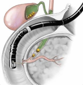
3. Olympus ERCP scope

4. state of the art operating theater with advanced instruments

5. harmonic scalpel

6. advanced bipolar for hemostatic control

7 . high definition camera for laparoscopic surgery

  • 1288 stryker camera
  • 1588 stryker camera

Faculty of gastro-enterology

  • Dr S B Das Chakroborty MD DM Gastroenterology
Faculty of gastrosurgery

·       Dr Avishek Mukherjee MS Surgery AIIMS MCh AIIMS

·       Dr Sanjoy Mondal MS AIIMS MCH AIIMS

Laparoscopic gall stone surgery


What is the gall bladder?

The liver is the chemical factory of the body. All the blood flows through the liver which then maintains the internal environment of the body. Liver produces the bile which collects within the liver and then flows out of it via the common bile duct.

The common bile duct drains into the duodenum, which is the first part of the small intestine. When a person is fasting then the extra bile produced by the person is stored inside the gall bladder. This is a side outpouching and it drains into the common bile duct, via a narrow connecting tube the cystic duct.

When the bile stagnates in the gall bladder it produces stones.

The gall bladder is a redundant organ of the body and contributes nothing to the digestive process but can be a source of significant morbidity and disease.

What are gall stones?


Gall stones are formed from stagnation of bile. There are two varieties of gall stones. Bile pigment stones and cholesterol accumulation stones. Gall stones are commoner among women in their forties.

What are the symptoms of gall stone disease?

Gall stones can be asymptomatic in a large proportion of patients and can be detected very often by routine ultrasonographic evaluation. However they can cause gall bladder colic by causing obstruction to the outflow of the bile.  This pain is quite severe and is located in the right upper part of the abdomen.

If the gall bladder gets infected then it can cause cholecystitis and in very advanced cases gangrene can also set in the gall bladder.

If the gall stones descend into the common bile duct and cause obstruction of the bile flow then the patient can develop obstructive jaundice.

What is the investigation needed for gall stone disease?


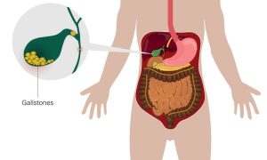
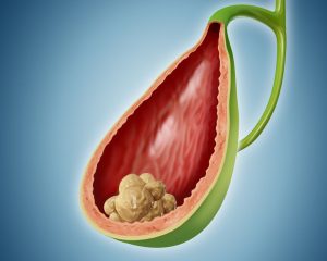
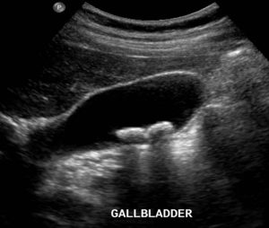
Gall stones are best detected by ultrasonography. This simple and painless test is very sensitive to detect gall stones and also assess the status of the common bile duct. We also will ask for routine liver function test and amylase lipase to assess the pancreatic function as well.

If  common bile duct stone or obstruction is suspected then we usually advise for an MRCP test which can outline the common bile duct better and more accurately.

How do we treat gall stone disease?


Gall stones If asymptomatic can be observed till they are symptomatic. But if the patient is diabetic then even asymptomatic stones should be treated since diabetics have a very high chance of complications like infection.

The best and only treatment for gall stone disease is surgical removal of the gall bladder. Although earlier open surgery was the norm now laparoscopic surgery has rather replaced open surgery.

The surgery is an excellent procedure wherein the gall bladder is removed after the cystic artery and the cystic duct are ligated.

The patient usually recovers very well and can return to normal life very fast.

What is the recovery period needed for gall stone disease surgery?

The patient is usually discharged after over night stay and can then take normal bath after one more day. The patient can move and walk from day one and after seven days can resume normal life and work.

Laparoscopic bile duct surgery


If the common bile duct is obstructed by stones, then we are able to free the stones from the common bile duct by laparoscopic exploration of the common bile duct. This surgery means that the stones can be removed from the common bile duct and the patient can be rid of obstructive jaundice while avoiding the need for open surgery.

Endoscopic retrograde cholangiopancreatography ERCP


When the common bile duct has stone induced obstruction we can clear the stones and place a stent to relieve the obstruction using endoscopic surgery.in ERCP we sedate the patient so the patient has no pain. Then we place an endoscope till the duodenum and observe the sphincter of oddi, this is the sphincter via which biliary duct and the pancreatic duct drain into the small intestine. Then we place an incision, opening this sphincter and then place a balloon catheter to remove the stones from within the bile duct. Subsequently a stent is usually  placed to allow the bile to drain and the patient to recover fast.

Laparoscopic hernia surgery What are hernias?


Hernia gaps or tears in muscles of the abdomen wall. When the tear develops then the content of the abdomen, mainly the intestine start protruding out of the gap and can often be felt as a swelling, like a small ball, which appear when the patient coughs or strains and then again reduces when the patient lies down and relaxes. In more advanced and neglected cases the intestine can get stuck inside the hernia sac wherein the patient may develop pain and then later symptoms of obstruction of the intestine.

How are they diagnosed?


Hernias are best diagnosed by clinical examination by a surgeon. However in case of any doubt then CT scan can very accurately diagnose the hernia.

Why should they be treated?

Hernias are best treated by surgery. Earlier open surgery was the normal, nowadays we treat them by laparoscopic surgery. And the results are excellent.

Are there any medicines or belts which can be used to treat hernias?

There are no medicines which can repair a gap in the abdominal muscle wall. Therefore hernias always require surgery to repair them. Belts which press the hernia opening can cause serious and sever damage to the intestine and therefore should never be used.

How do we treat them?


We treat all hernias by surgery, and we prefer laparoscopic surgery since the patient can recover fast after this surgery. The concept is we place a open port and then a camera into the abdomen. Generally first we reduce the content of the hernia, as in replace the content intestine, into the abdomen. Then we can define the extent of the muscle defect. Then we repair the defect in the muscle using a permanent repair.

What are mesh and what is darning repair of hernia?

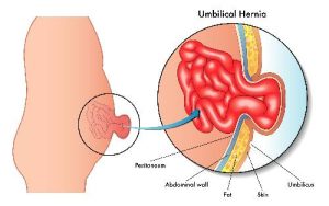
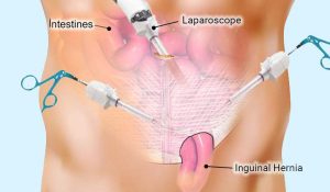
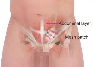
Since the muscle has a defect then we are able to fill the gap in the muscle by two techniques. Either we use a mesh, this is a synthetic cloth like material which is permanent in nature therefore it will permanently reinforce the gap in the muscle.

We can also join the gap in the musculature using a permanent suture made of polypropylene. The surgeon knits a loose mesh, like darning the muscle.

This repair I prefer since the mesh knitted by the surgeon is relaxed and tension free. Therefore, the patient has lesser pain. Also, the mesh can serve as a foreign body wherein if it ever gets infected, it creates a long standing and intractable infection. The mesh can also erode the bowel and cause life threatening complications.

Therefore, to avoid all these, we prefer the darning repair over the mesh repair of the hernias.

What is the recovery period of this surgery?

Generally, the patient recovers very smartly from this surgery, and the patient can resume normal life within seven days after the surgery

Make An Appointment