NKPC Kidney Clinic

Welcome to NKPC Kidney Clinic

Our urosurgical team

We have one of the largest and most experienced uro surgical team for the management of stone diseases. And therefore we are able to deliver best results in this disease care.

Dr Avishek Mukherjee MS AIIMS MCH PGI chandigarh

Head of uro-surgery, cancer surgery, and transplant surgery

Professor Dr Moloy Bera MS MCh

Senior consultant uro and cancer surgery

Dr Sandip Pramanik MS MCH

Senior consultant uro and cancer surgery

Dr Anurag Chaterjee MS MCH

Consultant uro surgery and cancer surgery


Dr Anand Biyani MS MCH

Consultant uro surgery and cancer surgery


Equipment for advanced therapy of renal stone disease Diagnostic

Ultrasound accuson from siemens Germany

32 slice CT scan siemens somatom

Auriga XL

Laser for advanced stone surgery


Advanced endosurgery equipment for the treatment of ureteral and renal stones

Ureteroscope Olympus Japan

Flexible uretero-renoscope Olympus Japan

Miniperc Olympus Japan

Pcnl nephroscope wolf surgical Germany

The kidney produce urine by filtration of the blood. The basic filtration unit of the kidney is a nephron. Each kidney has about ten lakh nephrons. These nephrons produce the urine and then drain into the calyces of the kidney. These calyces then drain into the pelvis of the kidney. The pelvis of the kidney drains the urine into the ureter. Each kidney has one ureter which drains the urine into the urinary bladder. The bladder then drains the urine via the urethra to outside the body. The prostate is located at the base of the bladder and it surrounds the urine exit route in males only. Females do not have prostate since the prostate in the males is replaced by the uterus in the females.

Why are stones formed?

Stones are formed in the urine by crystal formation of calcium oxalate in the urine. The urine also contains citric acid, which keeps the calcium in the urine dissolved, however when there is lesser quantity of citric acid in the urine of some people then the calcium crystalizes and stones are formed. These crystals then slowly grow in size with accumulation of more calcium.


How can they cause damage to the kidney?

Once the stones are formed and are small, they cause little damage. However as they grow larger then they can cause infection and obstruction to the flow of the urine therefore causing damage to the kidneys.


What are the symptoms of stone disease?

When stones are small and located in the calyces they hardly cause any pain or infection and generally silent. But as they grow larger and cause obstruction and infection then the patient becomes symptomatic. The classical presentation of kidney stones is pain. This pain is usually located in the side and flank area of the patient and radiates down to the lower abdomen. The pain can vary in severity from mild ache to severe pain.

When the stone is located in the renal pelvis or the ureter it causes obstruction and then the patient experiences colicky pain. This pain is very severe and radiates from the flank to the lower abdomen and can also radiate to the testis in men and to the vagina in females. This pain typically come and goes in waves.

Some patients can present with fever and infection. And in some cases. red colored urine, caused by bleed in the urine can also be a presentation. However this bleed is usually accompanied by pain which is colicky in nature.

What are the diagnostic test used to diagnose stone disease?

Case for 2weeks medical Trial

Case for Surgical Managment

When the patient presents to the uro-surgeon with symptoms suggestive of stone disease, he will generally advise for an ultrasound test but nowadays non contrast of plain CT scan has become the gold standard of detection of renal and ureteral stones.

In addition routine tests like creatinine hemoglobin and urine routine and culture are also ordered.

Ureteral stones

These are stones which have been formed in the kidney and have then descended into the ureter. Here they can cause severe pain and even damage to the kidneys by causing obstruction. Generally ureter stones are symptomatic, by causing severe pain and colic, but after some period when the kidney goes into shock then the stones may become silent. However the damage to the kidneys is ongoing.


Which ureteral stones can be treated medically?

When the ureteral stones are smaller than 5mm and are located in the lower part of the ureter, and the patient has not much pain, also the kidney is functioning well. These patients can be treated with medical management and observation.

Criteria for medical management of ureteral stones

Stones which are smaller than 5mm

Which are located in the lower part of the ureter

When the patient has not much pain

When the patient has no infection

When the patient is not diabetic

These patients can be selected for medical management of ureter stones.

Which patients should not be managed by medical management?

Any stones larger than 6mm are unlikely to pass by medicines and therefore are not selected for medical management.

Stones located in the upper part of the ureter are not likely to pass since the ureter becomes narrower as it travels towards the bladder therefore stones which are stuck in the wider upper part of the ureter are not likely to pass with medicines.

Stone which are causing severe pain  and infection should be immediately treated with surgery, or else the renal unit may get damaged.

Diabetic patients with poor sugar control are likely to get infected therefore diabetic patients are generally not selected for medical management.


How are these stones managed?

We generally use drugs like tamsulosin, these drugs dilate the lower ureter muscles, where the ureter enters the bladder. This is the narrowest part of the ureter. And when this is dilated the chance of stone passage increases.

We can also administer a preventive antiseptic drug like nitrofurantoin with the tamsulosin.


What is the duration of the trial period?

Follow up of patients has shown that the kidney can withstand obstruction for a period of two weeks after this period however renal damage starts. Therefore we recommend medical trial of expulsion of stone for not more than 2 weeks. If after two weeks of adequate medication yet the stone remains impacted it is better to remove these stones by surgery.

Which stones in the ureter need surgery?

Stones which are larger than 5mm, or are located in the upper ureter and not passing downwards, or are causing severe pain or infection, especially in a diabetic person, are better treated by primary endosurgical removal.

How are ureter stones operated?

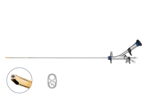
Open surgery for ureter stones has become obsolete and nowadays most stones are operated by endo-surgery. Endosurgery means operating using telescopes which can be introduced within the natural urinary passages to operate and remove ureteral stones.

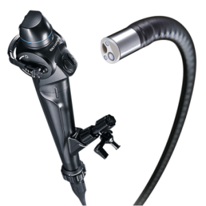
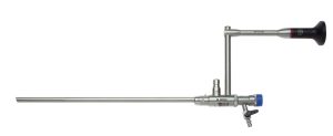
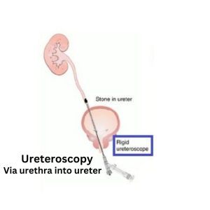
When stones are located in the lower ureter we operate using a telescope called the ureteroscope. And in upper ureter we use the flexible uretero-reno-scope.

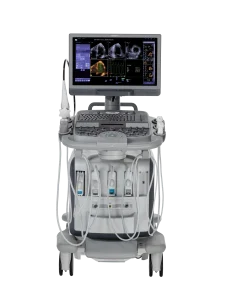
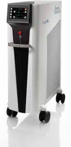
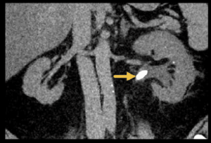
These telescopes allow us to access the ureter and visualize the stone. This endoscopic picture is seen on a medical grade high definition monitor. Which magnifies the picture 200 times. under this fine precision guidance we use the Auriga xl holmium laser to destroy the stone. The laser usually creates a plasma which destroys the stone by creating cavities within the stone. The stone often evaporates and very fine dust like particles remain. Which generally drain downstream with the flow of urine.

Management of stones located in the kidneys

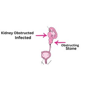
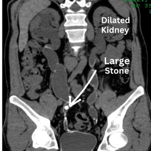
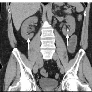
Stone which are located in the kidney also cause damage by obstruction and infection. And as such need to be treated too. There are no medications which can dissolve stones in the kidney or the ureter. Although lots of patients naturally desire this kind of stone dissolving therapy. This is yet not medically possible with current day medical care.

Which stones are insignificant stones?

Tiny stone non Obstructing Stone

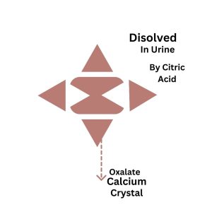
Stones which are smaller than 5mm and are not causing pain obstruction or infection can be treated as insignificant stones and need no further medical or surgical care. But to prevent growth of the stones we usually advise citric acid replacement therapy.

Citric acid is naturally available in citrus fruits and therefore we advise daily once intake of orange or musambi. These citrus fruits by increasing the levels of citric acid in the urine prevent growth of the stone.

Which stones need treatment?

Large obstructing stone,These need Surgery

Any stone larger than 5 mm and which is causing infection or pain and obstruction needs to be treated. Since there is no medical therapy therefore these stones are best treated by laser endosurgery. Open surgery and laparoscopic surgery are rarely needed for treatment of pure stone disease.

What is RIRS surgery?

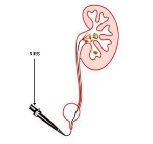
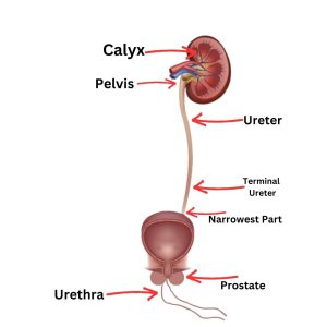
When the stone is located within the kidney and is smaller than 10mm in size it is best treated by RIRS. RIRS is retrograde intra-renal surgery. Herein we use a flexible uretero-renoscope. This is performed under general anesthesia, wherein the patient has no pain and the recovery is also very comfortable. The flexible scope is introduced via the external urinary opening and then travels via the bladder into the ureter and all the way into the renal calyces where these stones are destroyed by laser energy.

After this surgery generally a stent is placed which allows the kidney to heal and recover, and is generally removed after a period of two weeks to three months, depending on the extent of renal damage caused by the stone.

 What is miniperc surgery?

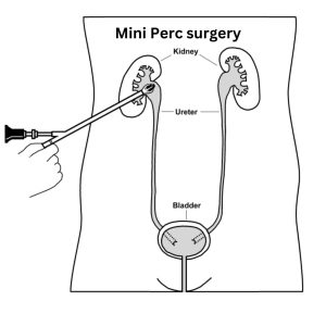
Miniperc Surgery,for stones between 10-20mm in the kidney or upper ureter

When the stone is larger than 10mm but smaller than 20 mmm it is best treated by miniperc surgery. This is performed also under general anesthesia. Herein a very tiny puncture is placed in the flank of the patient. And then we access the kidney using a mini nephroscope. Once the stone is located it is destroyed with laser energy. This is a highly effective treatment for larger kidney stones.

What is PCNL surgery?

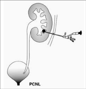
When the kidney has giant stones. Larger than 20mm in size then we use the large caliber nephroscope. These are larger caliber telescope which can visualize the stone and then remove larger stones from the kidney.

What are ureteral stents and when are they used?

Ureteral stent are needed when the ureter or kidney are dilated

Stent is placed from kidney into bladder

When the opening of the kidney pelvis into the ureter is edematous or swollen or the ureter is damaged or the kidney is dilated and needs time to recover, we may place a silicon rubber tube, from inside the kidney via the ureter into the urinary bladder. This helps the kidney to drain the urine and allows the kidney time to recover. After adequate recovery of the kidney the stent can then be removed. This period can vary from 2 weeks to more than 3-6 months. Depending on the nature of surgery performed and also the extent of damage of the kidney.

What is the typical recovery period after laser surgery for kidney stones?

Generally, the patients recover very well after the surgery. Typically, the patients have very little pain since the skin and muscles are hardly cut in these endosurgery. The patients are usually discharged between the second to the fourth post operative day. And can resume work after seven to fourteen days of rest at home.

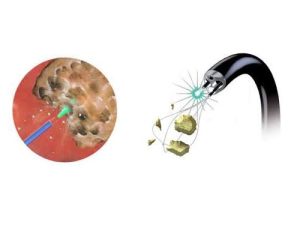
Laser creats a plasma bubble and destroys the stone


How to prevent recurrence of renal stones?

Since renal stones are formed due to the deficiency of citric acid in the urine therefore replacement of citric acid is the key to prevention of future reformation of stones. Citric acid is naturally available in citrus fruits and therefore we advise daily once intake of orange or musambi. These citrus fruits by increasing the levels of citric acid in the urine prevent growth of the stone.

We generally advise the patient to have one musambi or orange per day. Since the fruit pulp is also very valuable therefore one should chew and take the whole fruit while discarding the peel and the seeds. However if the serum creatinine is more than 2 and the kidneys are weakened then citrus fruits are best avoided to prevent hyperkalemia, increased levels of potassium in the blood.

In patients with multiple stones or history of recurrent stone we also add potassium citrate with magnesium citrate with vitamin B6, these are available as sugar free syrups and can be taken 5ml, 1 tea spoon, in one glass water, at bedtime lifelong, and to be stopped if kidneys become weaker in the future.

Make An Appointment